-
-
sample data

|
-
-
-
sample data

|
-
sample publication

|
|
FOO
Datasets
-> The Brain of the Dog in Section, Singer/Leedle
[ Canis lupus General
Info ]
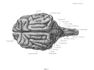
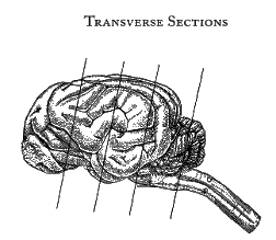
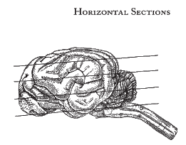
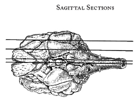
Surface drawings and transverse, horizontal, and sagittal sections of
the dog brain digitized from the out-of-print 1962 volume The
Anatomy of the Dog Brain in Section by Marcus Singer.
The following text is excerpted from A Digital Edition of the Brain
of the Dog in Section, available for download
here.
Preface to the digital edition
It is a great pleasure to bring
The Anatomy of the Dog Brain in Section by Marcus Singer
into the 21st century. The original
print edition was published in 1962, and has been out of print since
then . It is a work of extraordinary quality and remains the best
atlas of the canine brain anatomy available. Print copies are scarce
and they show their age. The dog brain atlas needed saving. With this
digital edition researchers and anatomists will no longer need to rely
on reference library copies or copies inherited from colleagues. No
one will need photocopies. Now anyone who “dumpster dives” to recover
a retiring colleague’s copy will do so only because he/she wishes an
original print edition and perhaps values the coffee stains which may
adorn it.
This digital edition began in 2000 as a scan of a library copy. At the
time, we were getting into neurotoxicology and needed neuroanatomy
references to locate specific brain regions, nuclei, and fiber
tracts. Like others, we were unable to purchase a canine neuroanatomy
text so we settled for a borrowed library copy of Dr. Singer’s
book. Luckily, we had an over-sized scanner available as the pages of
the original text are 12.25 x 14.5 inches in size.
Notes on the brains, plates, and labelling
The Brains
The sections reproduced in the book were obtained from the brains of
three disease-free beagle dogs of the same litter, aged five months,
raised at the Virus Research Laboratories of Cornell University.
Each brain was sectioned in one of the three planes: horizontal,
sagittal, and transverse. The dogs were anesthetized by
intraperitoneal injection of Nembutal solution and then were perfused
through the heart with a physiological saline solution followed by a
solution of 10 per cent formalin. The brains then were removed,
immersed in a solution of the same strength of formalin for a few
weeks, dehydrated in graded alcohols, and slowly infiltrated with
celloidin. The embedded brains were cut at 35 microns and the sections
numbered consecutively. Selected sections were stained by the
iron-hematoxylin method of Loyez for myelin sheaths after mordanting
in alum of iron (Bertrand). Adjacent sections, which alternated with
those stained for fibers, were stained for nerve cell bodies according
to the cresyl violet method of Bielschowsky-Plien (Bertrand). Loyez
sections are more useful for studying overall detail than sections
stained by the Nissl method, and the present atlas is confined to
sections stained for fibers. Indeed, nuclei, for example, of the
thalamus often can be distinguished more clearly in such
sections. However, constant reference has been made to sections of the
cresyl violet series in locating nuclear groupings in Loyez sections.
During dehydration and embedding, the brains shrank to about
three-fourths to two-thirds of the size of the fixed brain. The
sections were numbered consecutively: the sagittal sections, from left
to right; the horizontal sections, from ventral to dorsal; and the
transverse sections, from rostral to caudal. The orientation of the
sections corresponded closely to the desired plane. However, the
sagittal sections were tilted about 1 mm from the true plane so that
the midline section lies approximately in section number 575 (plate
59) for the medulla but in about section 605 for the rostral part of
the brain stem. The transverse plane corresponded to a cut from the
posterior commissure to the caudal extremity of the mamillary bodies
(see transverse section 826, plate 35). Since the brain stem of the
dog, unlike that of the human, is flexed only slightly at the
mesencephalon, reorientation of the embedded specimen was not
required, and the sections of the upper and lower brain stem deviated
only slightly in direction from one another. The bilateral orientation
of the transverse sections diverged only a little from the true
orientation, the left being slightly more rostral than the right. In
the case of the horizontal sections, a plane was chosen cutting
through the anterior commissure rostrally and the decussation of the
trochlear nerve caudally (see section 383, plate 114, Commissura
anterior and Decussatio nervorum trochlearium). The bilateral
orientation of the horizontal plane was tilted slightly so that the
left side was cut a little more deeply than the right (for example,
see section 456, plate 110, Nucleus corporis geniculati).
The Selection of Sections
Sections spaced at suitable intervals were selected for full
reproduction in the atlas. The selection included 49 transverse
sections, 27 sagittal sections, and 28 horizontal ones. In addition,
15 of the most medially placed sagittal sections were further enlarged
and the enlargements are reproduced in the book. Consequently, the
atlas consists of 119 plates of sections and 5 drawings of surface
topography.
In the sagittal series, successive sections are separated in most
cases by an interval of 10 (approximately 350 microns) except in
certain regions. The most medial section is numbered 595 (plate 55)
and the most lateral, 265 (plate 96). The spacing permitted
visualization of all major structures. In the exceptions, the interval
of separation between successive sections was reduced or increased. In
the former instance, the interval from 475 (plate 83) to 485 (plate
77) also included sections 478 (plate 81) and 482 (plate 79) in order
to show rapidly changing detail and to label the great profusion of
structure that could not be accommodated in fewer sections. In the
latter case, the most lateral sections, which showed little change in
the 10 sections lateral to section 435 (plate 88), generally were
spaced at intervals of 30 and 20 sections. In the enlargements of the
15 most medial sections, the brain stem was magnified further and
cortical structures were largely omitted, a procedure adopted
previously in the atlas of sagittal sections of the human brain
(Singer and Yakovlev). The enlarged photographs of the brain stem
served to clarify some of the detail and to multiply the space for
labeling. Such special enlargements were employed for slides 595
(plate 55) to 475 (plate 83), inclusive.
The horizontal sections were also arranged in intervals of every tenth
section, except in the most dorsal and ventral regions where the
spacing was increased. In some instances, for one reason or another,
the section was inadequate for reproduction, and therefore an adjacent
one was substituted, thus introducing a disparity in the number
sequence.
In the case of the transverse sections, the spacing between successive
sections is in most instances about 20. In a few instances, the
interval was less and at the rostral or caudal extremes was as great
as 40 or 50.
The serial number of the section is recorded at the top right of each
plate. Consequently, the distance of each section from any other of the
same plane can be calculated by the reader, since each section is
about 35 microns in thickness. For example, in the sagittal series,
the interval between section 465 (plate 85) and the midline section
595 (plate 55) is 130 times 35 microns or approximately 4.5 mm.
Labeling Techniques
All labeling was done on a transparent paper overlay and then
transferred in final form to a cellulose acetate overlay. The
structures were labeled directly with lead lines, and the terms were
abbreviated in a standard and simple way. The method of direct
labeling was used in preference to symbols in order to preserve the
beauty of the sections and to facilitate the identification of
structures by the reader. Because of the wealth of structure to be
identified, this method precluded complete labeling of each
section. Consequently, most structures are not identified on every
plate in which they occur but instead are labeled on alternate or
occasional plates.
When a structure was labeled on a number of plates in one of the
planes of section, the label generally was placed in a similar
position on each plate for the convenience of the reader.
Finally, it should be noted again that the sagittal sections were
taken from the left side. However, in most medial sections the caudal
part of the brain stem included part of the right side because of the
orientation of the block during sectioning. Whenever right-sided
structures are shown, (R) is affixed to the term.
|
|